
Cell Viability with MTT Assay Summary
Cell Viability is a common technique used by biochemists who are studying oncology and pharmaceutics. The most common use for cell viability studies is when determining the IC50 for a cytotoxic compound in cell culture. However, as you can expect, there are a lot of different times when you need to know if your cells are alive. In larger pharmaceutical companies, MTT Cell Viability studies for Cytotoxic compounds are performed as a high throughput method because companies routinely screen MASSIVE libraries of small molecule drugs. To measure cell viability, researchers typically use an MTT assay, Cell Titer Blue, Trypan blue exclusion, or ATP assay. In this method guide, we will walk through the theory behind all these methods and then end with a protocol for the MTT assay. It would be a great test of your skills if you could use our High Performance Liquid Chromatography (HPLC) Method Guide to detect the products of the MTT assay.
Note: If you’re writing research papers, I highly recommend Grammarly – it’s a free grammar check plugin for Chrome. Try it out here…
Using an MTT Assay to measure Cytotoxicity
In general, to measure cell viability, you need to incubate cells with a reagent and measure the conversion of your reagent into a product. If lots of cells are alive, most of your reagent will be converted. If lots of cells are dead, then your reagent will only be partially converted. For the MTT assay, the reagent used is
(3-(4,5-dimethylthiazol-2-yl)-2,5-diphenyltetrazolium bromide) tetrazolium. This is a positively charged small molecule that undergoes NADPH-mediated conversion over to Formazan. Because of its positive charge, MTT can enter viable cells and non-viable cells with ease. Upon conversion, the Formazan product precipitates inside cells near the cell surface and can be detected using a spectrophotometer. Note: MTT only needs an intact and functioning mitochondria to be converted so it is a metabolic assay and not a proliferation assay. I’ll discuss cell proliferation assays in the future. The assay technique is very simple:
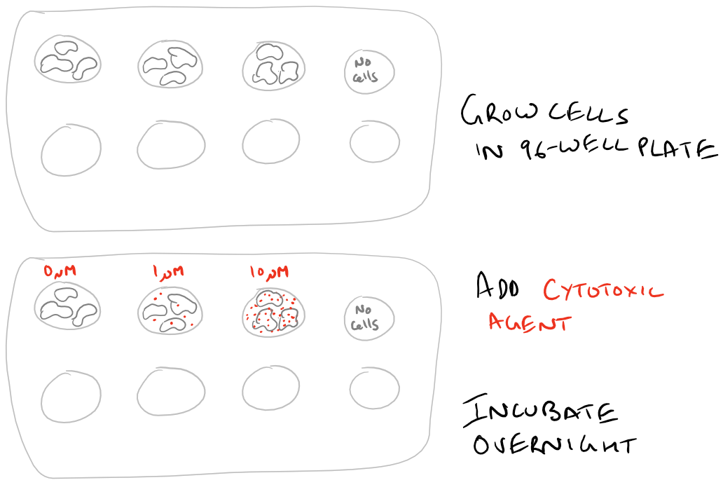
- Grow an equal number of cells in different wells of a microplate
- Add your cytotoxic compound and incubate
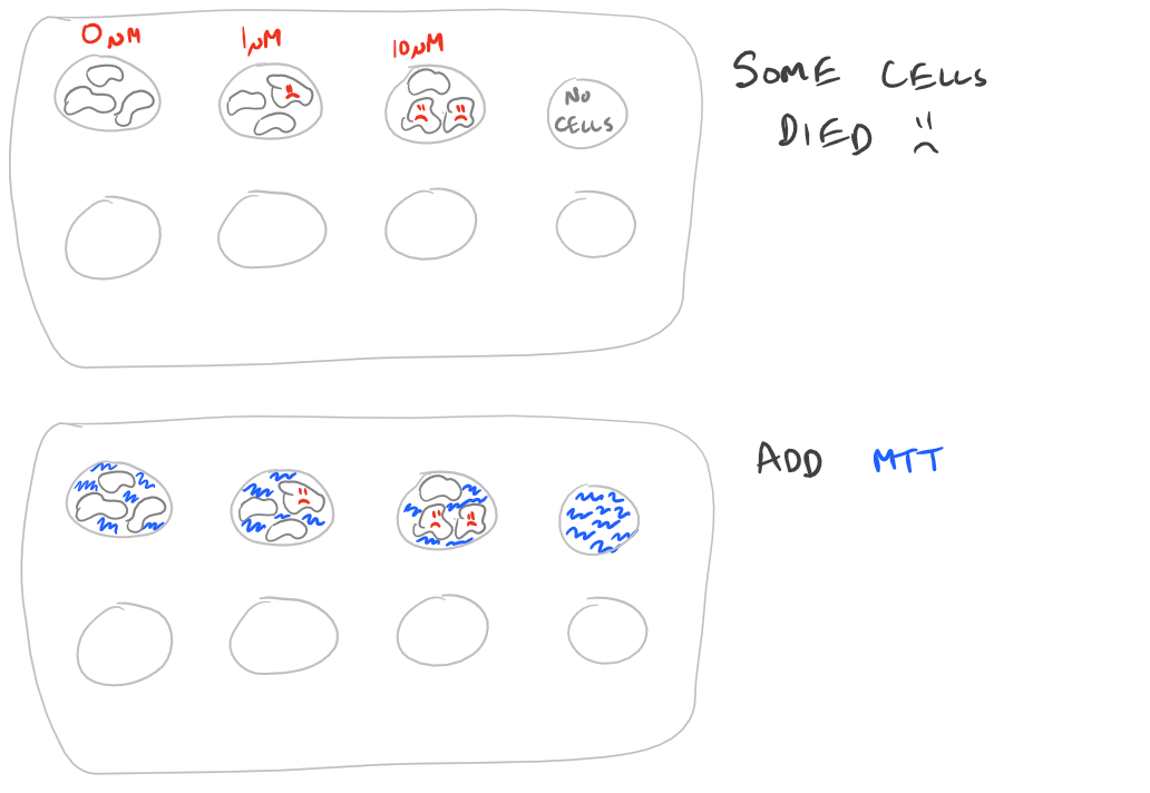
- Then replace the media and add the MTT, let the cells convert the MTT (blue) into Formazan (purple)
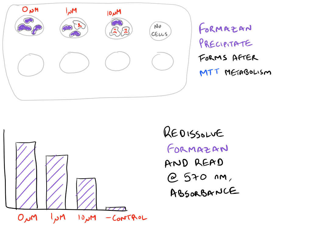
- Use SDS along with DMF or DMSO to resolubilize the formazan and to kill cells (stop them from converting any more reagent)
- Then measure how much formazan was created using a spectrophotometer.
Take a look below to understand these steps:
Some researchers have even combined the use of the MTT assay with Flow Cytometry (FACS) to sort viable cells from non-viable cells. However, this is uncommon and there are much better stains for FACS such as Propidium iodide (PI).
Cell Titer Blue, Trypan Blue and ATP Assays
As noted above, the MTT assay is really a metabolic assay because the MTT molecule needs to enter a cell and get converted to Formazan using NADPH. While the exact mechanism of MTT’s metabolism isn’t clear, this means the mitochondria needs to be intact and functioning. So, if you add a cytotoxic material which reduces mitochondrial efficiency, you might get weird results. In this case, it’s useful to also know other live/dead assays. The other major cell viability assays that are used in research include:
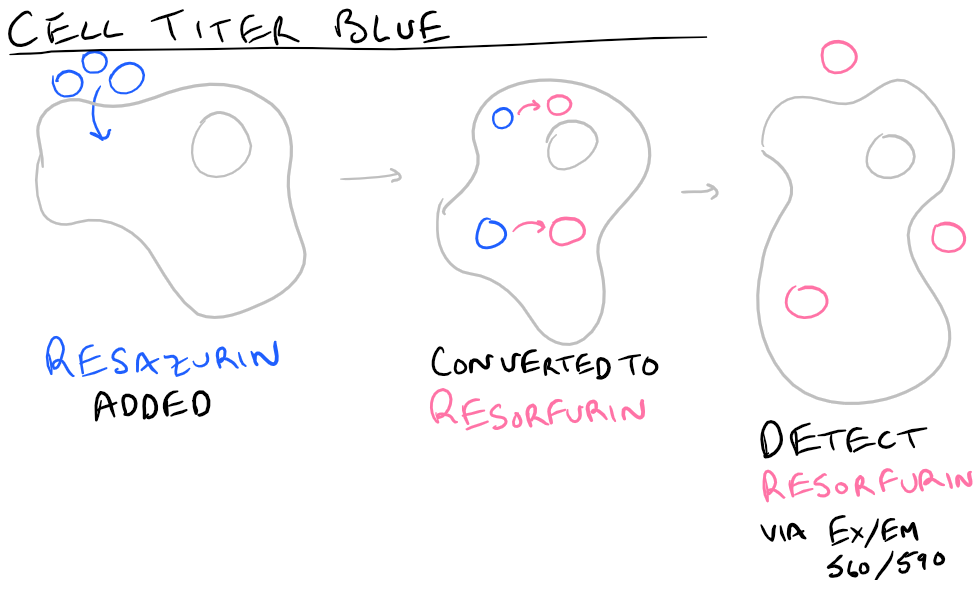
Cell Titer Blue: Similar to the MTT Assay, this assay involves incubating cells with resazurin (blue) and forming resorfurin (pink) after the cells metabolize it. Generally the metabolism takes 1-4 hours but it is much more sensitive than the MTT assay because you can measure the product via fluorescence (Ex/Em 560 nm/590 nm). The main advantage of this assay is that you don’t need to resolubilize the product in DMF/SDS so it’s much simpler. This is also a great high throughput assay!
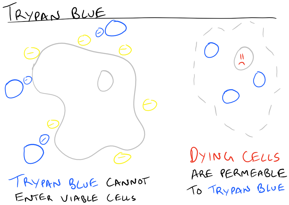
Trypan Blue Exclusion Assay: If you don’t have a spectrophotometer, then it’s simple to use the trypan blue staining method along with a microscope. Because trypan blue is a charged dye, it cannot permeate through living cells. So, simply incubating cells with trypan blue and looking under a microscope allows you to visually determine the # of viable cells (unlabeled), # of non-viable cells (blue), and the # of damaged cells (slightly blue). Count the number of cells in different fields of view and you’re done! Viability is just the ratio of live cells divided by total number of cells. The disadvantage with this method is that all you test is the membrane integrity of the cells. You don’t know if the cells are truly non-viable or just damaged a little bit.
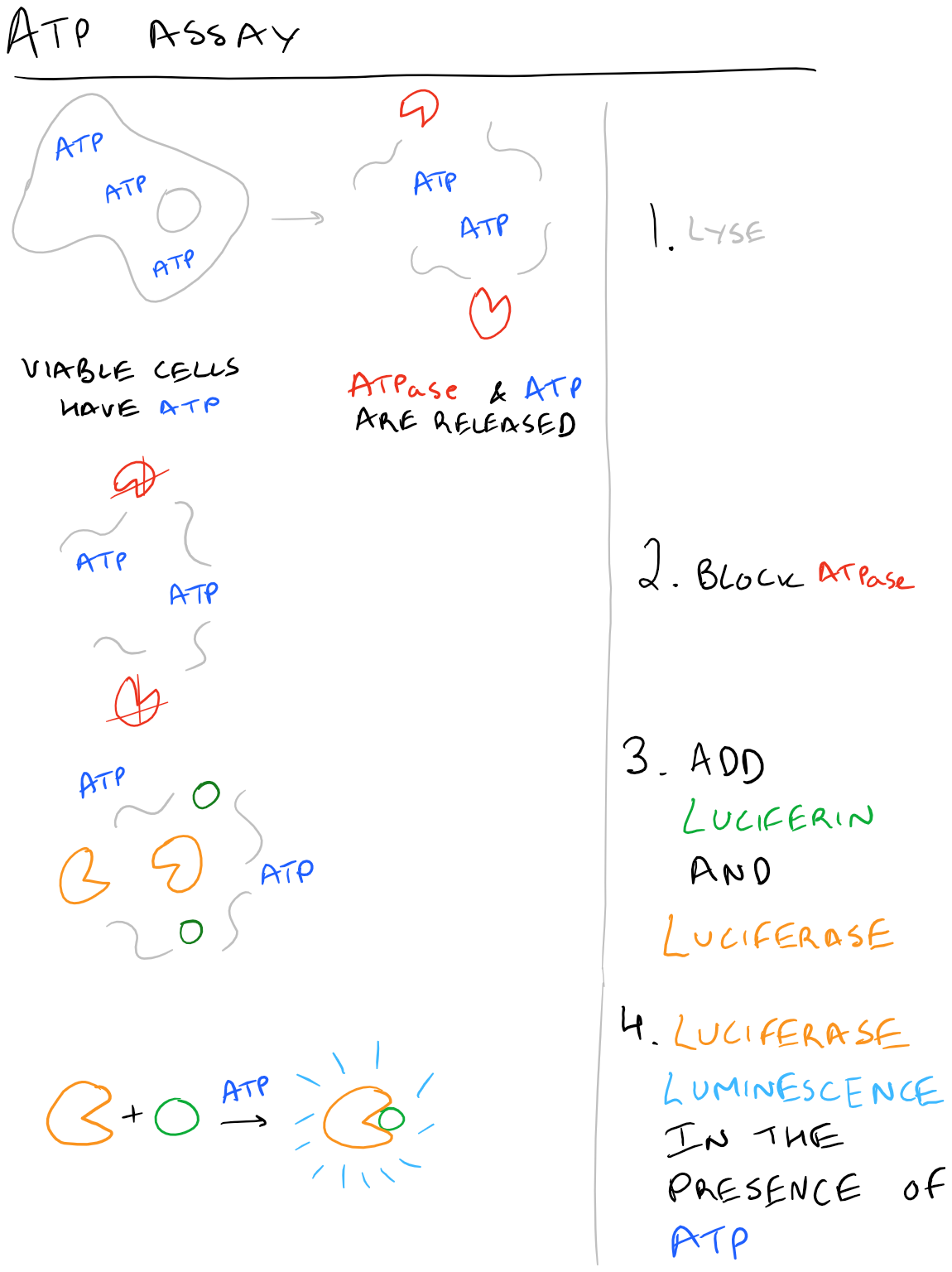
ATP Assays: When cells are non-viable, they cannot make any more ATP whereas viable and happy cells can make ATP. Additionally, as soon as cells die, ATPases rapidly break down ATP. Using these bits of information, it’s easy to see why an ATP based assay would work really well. The theory is simple – lyse cells, stop ATPases from hydrolyzing ATP, add in Luciferase and Luciferin. You’ll get excellent luminescence signal for hours!
Note: there are several other MTT-like molecules which are also used in cell viability assays: MTS, XTT, WST-1. The general principle however is all the same. The only note-worthy difference is that some of these molecules don’t penetrate live-cells, so they give you the reverse signal (how many dead cells there are).
Here are some images describing the above methods:
Side-note: Use Grammarly to check your research papers – it’s a free grammar check plugin for Chrome. Try it out here…
Cell Viability with MTT Assay Protocol
Materials for MTT Assay
MTT Solution (5 mg/ml MTT in PBS, pH 7.4, #M2128 Sigma Aldrich)
Solubilization solution, recipe here:
- 40% v/v Dimethylformamide #D4551 Sigma Aldrich
- 2% Glacial Acetic Acid #320099 Sigma Aldrich
- 16% Sodium Dodecyl Sulfate #436143 Sigma Aldrich
- pH 4.7 & 37oC
96 well plate
Hep G2 cells
Complete DMEM (indicator-free, no phenol-red) with 10% Fetal Bovine Serum
PBS
Cytotoxic compound (ex: Doxorubicin)
Step-by-Step Cell Viability MTT Assay
- Make the above solutions. Store MTT solution protected from light at 4oC and make sure there is no precipitate in the Solubilization solution.
- Seed 25 x 103 Hep G2 cells in a 96 well plate with 250 ul of DMEM.
- Add your cytotoxic compound (5 uM for Doxorubicin). Incubate for a desired time period (24 hours for Doxorubicin).
- Aspirate media and wash 3x with PBS.
- Add 125 ul of DMEM with 25 ul of MTT Solution. Incubate for 2 hours at 37oC.
- Add in 100 ul of solubilization solution.
- Pipette gently to mix without creating bubbles.
- Measure via absorbance at 570 nm using spectrophotometer.
MTT Assay Notes, Tips, and Tricks
- Always set up positive and negative controls! For positive controls have cells untreated with any cytotoxic compound as part of your wells. For negative controls have cells treated with 3% SDS as part of your wells. Also, make sure to have wells that have no cells, only media.
- Increasing the number of cells also increases your signal
- Too much MTT forms Formazan crystals which will damage cells so you might see the cells changing morphology.
- This is an end-point assay because the precipitate inside the cells will kill them. Don’t plan on keeping your cells alive for any further studies after you add the MTT.
- Having thiol-containing compounds in solution will convert MTT over to Formazan, so you’ll get false-positive data.
- Having phenol-red in your medium may also convolute your results. Dye-free media is important to use.
Cell Viability Protocols on SciGine
- Cell Viability of A540 cells treated with Gleevec (Cytotoxic agent) using MTT Assay
- Cell Viability after infection with HIV-1
- Human Pancreatic Cancer Cell Viability with MTT Assay
- Cytotoxic agent Docosahexanoic acid prevents KPL-1 cancer growth, detected via MTT assay
- Apoptosis during Myotube formation detected via Cell Titer blue and Trypan Blue exclusion
Checking IC50/Cell Viability via MTT Assay with Cytotoxic compound: Video
References
References
Excellent NIH guide related to Cell Viability
Trypan Blue Exclusion information
Discussing effectiveness of MTT assay for leukemia research
[…] the theory behind these assays and how to apply them in the lab. Combining our techniques of MTT Cell Viability assays and Flow Cytometry or FACS, we are really building up a great list of skills to analyze biological […]